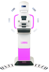
Designed to meet the needs of modern medical practices, the PANOX Digital Mobile X-ray HIGHPLUS combines state-of-the-art imaging capabilities in a portable solution.
The Panox ORIENT Digital Mobile X-ray Engineered with only 580mm width to Move along narrowest spaces freely, the system designed to ensure clear visibility view to during transportation of the device.
The Panox ORIENT Digital Mobile X-ray Low dose exposure mode significantly minimize the clinical dosage. And achieves sharp detailed medical images
This innovative feature allows the device to rotate a full 315 degrees, providing imaging access to even the most challenging anatomical positions and ensures accurate and precise diagnostic imaging
PALUS digital x ray detectors switching in one button from dynamic to static, quickly meeting patients image acquisitions needs.
PALUS digital x ray is equipped with “10” touchscreen display reviewing patient’s info and displaying exposure parameters control.
The collimation field will be set automatically according to chosen parameters reducing diagnostic workload time. Auto-tracking between X-ray tube and detector Auto-alignment between tube and detector offering accurate positioning for image acquisition.
Wireless _at panel detectors, using thick cesium iodide with high DQE to show tiniest details of bones and tissues for accurate diagnosis 14×17 – 17x 17 Si/Csi wireless, High FPD providing accurate acquisition of every image detail
Examination revolution decreasing the time of exam from 3 Minutes to only 40 Seconds
Lorem Ipsum is simply dummy text of the printing and typesetting industry. Lorem Ipsum has been the industry’s standard dummy text ever since the 1500s, when an unknown printer took a galley of type and scrambled it to make a type specimen book. It has survived not only five centuries, but also the leap into electronic typesetting, remaining essentially unchanged. It was popularised in the 1960s with the release of Letraset sheets containing Lorem Ipsum passages, and more recently with desktop publishing software like Aldus PageMaker including versions of Lorem Ipsum.
PALUS digital x ray detectors switching in one button from dynamic to static, quickly meeting patients image acquisitions needs.
PALUS digital x ray is equipped with “10” touchscreen display reviewing patient’s info and displaying exposure parameters control.
The collimation field will be set automatically according to chosen parameters reducing diagnostic workload time. Auto-tracking between X-ray tube and detector Auto-alignment between tube and detector offering accurate positioning for image acquisition.
Wireless _at panel detectors, using thick cesium iodide with high DQE to show tiniest details of bones and tissues for accurate diagnosis 14×17 – 17x 17 Si/Csi wireless, High FPD providing accurate acquisition of every image detail
Examination revolution decreasing the time of exam from 3 Minutes to only 40 Seconds